
Dosya:Schistosoma haematobium egg 4843 lores.jpg

Daha yüksek çözünürlüğe sahip sürüm bulunmamaktadır.
Schistosoma_haematobium_egg_4843_lores.jpg ((700 × 460 piksel, dosya boyutu: 38 KB, MIME tipi: image/jpeg))
Bu dosya Wikimedia Commons'ta bulunmaktadır. Dosyanın açıklaması aşağıda gösterilmiştir. Commons, serbest/özgür telifli medya dosyalarının bulundurulduğu depodur. Siz de yardım edebilirsiniz. |
| Açıklama |
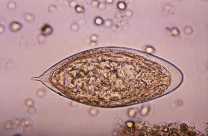
ID#:4843 This micrograph depicts an egg from a Schistosoma haematobium trematode parasite; magnified 500x. Note the egg's posteriorly-protruding, terminal spine, unlike the spinal remnant, which protrudes from the lateral wall of the Schistosoma japonicum egg. These eggs are eliminated in an infected human's feces or urine, and under optimal conditions in a watery environment, the eggs hatch and release "miracidia", which then penetrate a specific snail intermediate host. Once inside the host, the S. haematobium parasite passes through two developmental generations of sporocysts, and are released by the snail into its environment as "cercariae". |
|||
| Tarih | ||||
| Kaynak | http://phil.cdc.gov/PHIL_Images/20031013/b47fc1793d7443d7a5cdbfbc73d95e53/4843_lores.jpg | |||
| Yazar | CDC, Public Health Image Library (PHIL) | |||
| İzin (Bu dosyanın tekrar kullanımı) |
|
Dosya geçmişi
Dosyanın herhangi bir zamandaki hâli için ilgili tarih/saat kısmına tıklayın.
| Tarih/Saat | Küçük resim | Boyutlar | Kullanıcı | Yorum | |
|---|---|---|---|---|---|
| güncel | 17.13, 7 Mayıs 2006 | | 700 × 460 (38 KB) | Patho | {{Information| |Description=ID#: 4843 Description: This micrograph depicts an egg from the trematode parasite Schistosoma japonicum with its vestigial spine. The Schistosoma japonicum egg is typically oval or subspherical, has a vestigial spine, and is |
Dosya kullanımı
Bu görüntü dosyasına bağlantısı olan sayfalar:
Küresel dosya kullanımı
Aşağıdaki diğer vikiler bu dosyayı kullanır:
- ar.wikipedia.org üzerinde kullanımı
- cs.wikipedia.org üzerinde kullanımı
- de.wikibooks.org üzerinde kullanımı
- en.wikipedia.org üzerinde kullanımı
- fr.wikipedia.org üzerinde kullanımı
- gl.wikipedia.org üzerinde kullanımı
- ha.wikipedia.org üzerinde kullanımı
- nl.wikipedia.org üzerinde kullanımı
- sw.wikipedia.org üzerinde kullanımı
- zh.wikipedia.org üzerinde kullanımı