
Dosya:Carboxysome 3 images.png

Bu önizlemenin boyutu: 800 × 369 piksel. Diğer çözünürlükler: 320 × 147 piksel | 640 × 295 piksel | 1.024 × 472 piksel | 1.280 × 590 piksel | 2.943 × 1.356 piksel.
Tam çözünürlük ((2.943 × 1.356 piksel, dosya boyutu: 3,72 MB, MIME tipi: image/png))
Bu dosya Wikimedia Commons'ta bulunmaktadır. Dosyanın açıklaması aşağıda gösterilmiştir. Commons, serbest/özgür telifli medya dosyalarının bulundurulduğu depodur. Siz de yardım edebilirsiniz. |
Özet
| Açıklama |
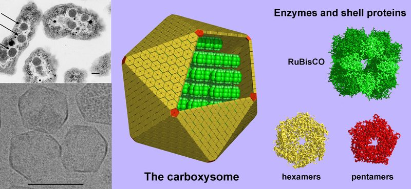
English: (Left, above) A thin-section electron micrograph of H. neapolitanus cells with carboxysomes inside. In one of the cells shown, arrows highlight the visible carboxysomes. (Left, below) Purified carboxysomes (material courtesy of S. Heinhorst and G. Cannon) as visualized by cryo-electron microscopy (courtesy of M. Yeager and K. Dryden). (right) Models for the structure of the carboxysome. Current data suggest that the shell is composed of several hundred hexameric protein building blocks and 12 pentameric building blocks. The three-dimensional atomic structures of the shell proteins have been determined by X-ray crystallography. RuBisCO, the main interior enzyme is shown packed inside in a regular arrangement for simplicity, though the actual organization of the enzymes is not understood yet. The other key enzyme, carbonic anhydrase, which is present in lesser amounts, is not illustrated. Scale bars are 100 nm. (image by T. Yeates). |
| Kaynak | Modified version of Image:Carboxysome.png incorporating image taken from Image:Carboxysomes EM.jpg |
| Yazar | Prof. Todd O. Yeates, UCLA Dept. of Chem. and Biochem. |
| Diğer sürümler |
[]
PNG:
|


Lisanslama
Bu dosya, Creative Commons Atıf 3.0 Uluslararası lisansı ile lisanslanmıştır
- Şu seçeneklerde özgürsünüz:
- paylaşım – eser paylaşımı, dağıtımı ve iletimi
- içeriği değiştirip uyarlama – eser adaptasyonu
- Aşağıdaki koşullar geçerli olacaktır:
- atıf – Esere yazar veya lisans sahibi tarafından belirtilen (ancak sizi ya da eseri kullanımınızı desteklediklerini ileri sürmeyecek bir) şekilde atıfta bulunmalısınız.
Dosya geçmişi
Dosyanın herhangi bir zamandaki hâli için ilgili tarih/saat kısmına tıklayın.
| Tarih/Saat | Küçük resim | Boyutlar | Kullanıcı | Yorum | |
|---|---|---|---|---|---|
| güncel | 01.38, 6 Ağustos 2008 | | 2.943 × 1.356 (3,72 MB) | TimVickers | {{Information |Description={{en|1=(Left, above) A thin-section electron micrograph of H. neapolitanus cells with carboxysomes inside. In one of the cells shown, arrows highlight the visible carboxysomes. (Left, below) Purified carboxysomes (material court |
Dosya kullanımı
Bu görüntü dosyasına bağlantısı olan sayfalar:
Küresel dosya kullanımı
Aşağıdaki diğer vikiler bu dosyayı kullanır:
- ar.wikipedia.org üzerinde kullanımı
- bg.wikipedia.org üzerinde kullanımı
- en.wikipedia.org üzerinde kullanımı
- en.wikiversity.org üzerinde kullanımı
- sw.wikipedia.org üzerinde kullanımı