
Dosya:Chlamydomonas TEM 09.jpg

Bu önizlemenin boyutu: 751 × 600 piksel. Diğer çözünürlükler: 301 × 240 piksel | 601 × 480 piksel | 961 × 768 piksel | 1.280 × 1.023 piksel | 1.800 × 1.438 piksel.
Tam çözünürlük ((1.800 × 1.438 piksel, dosya boyutu: 784 KB, MIME tipi: image/jpeg))
Bu dosya Wikimedia Commons'ta bulunmaktadır. Dosyanın açıklaması aşağıda gösterilmiştir. Commons, serbest/özgür telifli medya dosyalarının bulundurulduğu depodur. Siz de yardım edebilirsiniz. |
| Açıklama |
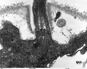
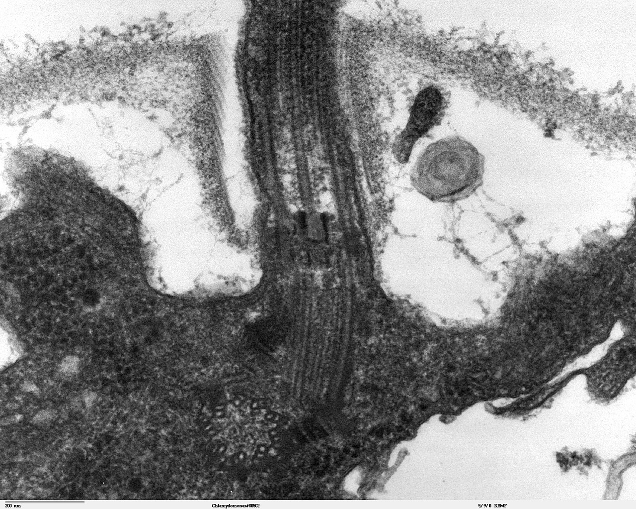
Transmission electron microscope image, showing an example of green algae (Chlorophyta). Chlamydomanas reinhardtii is a unicellular flagellate used as a model system in molecular genetics work and flagellar motility studies. This image is a longitudinal section through the flagella area. In the cell apex is the basal body that is the anchoring site for a flagella. Basal bodies originate from and have a substructure similar to that of centrioles, with nine peripheral microtubule triplets(see structure at bottom center of image). The two inner microtubules of each triplet in a basal body become the two outer doublets in the flagella. This image also shows the transition region, with its fibers of the stellate structure. The top of the image shows the flagella passing through the cell wall. |
| Tarih | |
| Kaynak | Source and public domain notice at http://remf.dartmouth.edu/imagesindex.html |
| Yazar | Dartmouth Electron Microscope Facility, Dartmouth College |
| İzin (Bu dosyanın tekrar kullanımı) |
Released into the public domain |
| Bu iş yazarı Dartmouth Electron Microscope Facility, Dartmouth College tarafından kamu malı olarak yayınlanmıştır. Bu dünya çapında geçerlidir. Bazı ülkelerde bu yasal olarak mümkün olmayabilir; öyleyse: Dartmouth Electron Microscope Facility, Dartmouth College, bu işi herhangi bir amaç için, herhangi bir şart olmaksızın, yasalarca gerekli olmadıkça, herkesin kullanmasına izin veriyor.
|
Dosya geçmişi
Dosyanın herhangi bir zamandaki hâli için ilgili tarih/saat kısmına tıklayın.
| Tarih/Saat | Küçük resim | Boyutlar | Kullanıcı | Yorum | |
|---|---|---|---|---|---|
| güncel | 06.47, 21 Eylül 2007 | | 1.800 × 1.438 (784 KB) | Neil916 | {{Information |Description= Transmission electron microscope image, showing an example of green algae (Chlorophyta). <br><br>''Chlamydomanas reinhardtii'' is a unicellular flagellate used as a model system in molecular genetics work and flagellar motilit |
Dosya kullanımı
Bu görüntü dosyasına bağlantısı olan sayfalar:
Küresel dosya kullanımı
Aşağıdaki diğer vikiler bu dosyayı kullanır:
- ar.wikipedia.org üzerinde kullanımı
- bs.wikipedia.org üzerinde kullanımı
- ca.wikipedia.org üzerinde kullanımı
- cs.wikipedia.org üzerinde kullanımı
- de.wikipedia.org üzerinde kullanımı
- de.wikibooks.org üzerinde kullanımı
- en.wikipedia.org üzerinde kullanımı
- es.wikipedia.org üzerinde kullanımı
- gl.wikipedia.org üzerinde kullanımı
- id.wikipedia.org üzerinde kullanımı
- ja.wikipedia.org üzerinde kullanımı
- ko.wikipedia.org üzerinde kullanımı
- pl.wikipedia.org üzerinde kullanımı
- ru.wikipedia.org üzerinde kullanımı
- sv.wikipedia.org üzerinde kullanımı
- uk.wikipedia.org üzerinde kullanımı
- zh.wikipedia.org üzerinde kullanımı